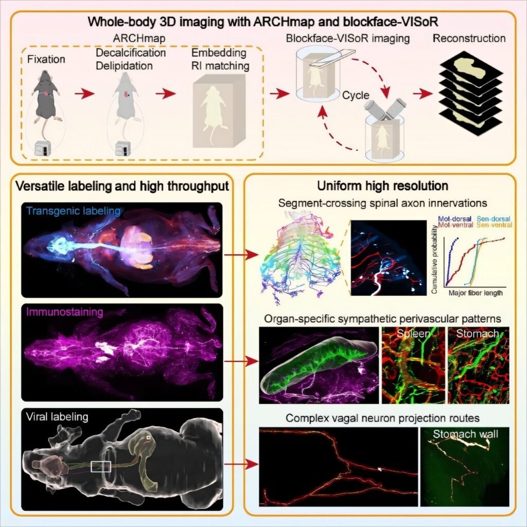
A groundbreaking advancement has been achieved in the realm of three-dimensional (3D) imaging of large-scale biological tissues. Scientists have successfully developed the fastest high-definition 3D imaging technology in the world, capable of capturing the entire body of small animals at subcellular resolution. This cutting-edge technology allows for the efficient mapping of the intricate architecture of the peripheral nervous system (PNS). The remarkable findings of this study were recently published in the prestigious scientific journal, Cell.
The research team, spearheaded by Professors Guo-Qiang Bi and Pak-Ming Lau from the University of Science and Technology of China (USTC), collaborated with the Institute of Artificial Intelligence at the Hefei Comprehensive National Science Center and the Shenzhen Institute of Advanced Technology, Chinese Academy of Sciences to achieve this remarkable feat.
The peripheral nervous system plays a vital role in mediating bidirectional communication between the brain and various organs in the body. It facilitates the transmission of motor commands to regulate essential functions like breathing and heartbeat, while also processing and relaying sensory signals such as pain and temperature. Understanding the complex functional mechanisms of the PNS and its role in disease pathogenesis necessitates a comprehensive mapping of its intricate connections throughout the entire body.
Traditionally, knowledge of PNS architecture has been limited to millimeter-resolution anatomical studies. While recent advancements in 3D optical microscopy have enabled micron-resolution mapping of the whole brain, similar analyses for the PNS on a whole-body scale have posed significant challenges.
Existing imaging techniques have struggled to balance resolution and speed when it comes to capturing the complex and intermingled nerve pathways of the PNS throughout the entire body. The team’s previous development of the Volumetric Imaging with Synchronized On-the-Fly Scan and Readout (VISoR) technology for 3D imaging of cleared brain samples paved the way for this groundbreaking achievement.
To address the challenges posed by imaging whole mouse bodies, the researchers pioneered a novel approach combining “in situ sectioning + 3D blockface imaging.” By integrating a precision vibratome and the ARCHmap protocol for whole-body clearing and hydrogel embedding, they developed a blockface-VISoR imaging system.
This innovative system captures 3D surface images at a depth of approximately 600 μm before automatically removing 400-μm-thick tissue slices in a cyclical manner. Seamless 3D alignment is achieved through automated inter-section stitching algorithms, utilizing overlapping regions between adjacent sections. The shallow imaging depth minimizes light scattering in cleared tissue, enabling high-resolution imaging.
Through this groundbreaking approach, the researchers successfully achieved uniform subcellular-resolution 3D imaging of an entire adult mouse body within 40 hours, generating a vast amount of data per fluorescence channel. This technology is compatible with common neuroscience labeling methods, allowing for detailed analysis of various peripheral nerves and their projection paths throughout the body.
The implications of this breakthrough extend beyond the realm of neural regulation, offering insights into developmental biology, comparative anatomy, and biomedical research. Future optimizations of this technology may involve multi-channel imaging and exploration of its application in imaging other large-scale biological samples.
In conclusion, the remarkable achievement of high-definition panoramic imaging of the entire mouse body represents a significant leap forward in our understanding of the peripheral nervous system and its intricate connections throughout the body. This groundbreaking technology opens new avenues for research and discovery in the field of neuroscience and beyond.


