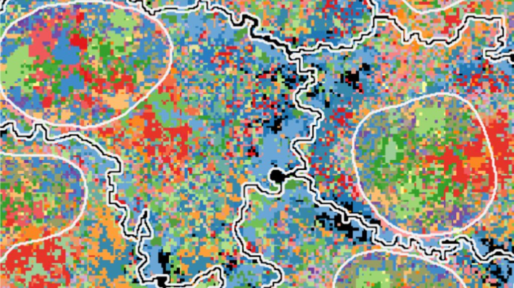
The field of microscopy has come a long way in helping researchers study diseases like cancer and improve therapies. Advanced instruments now allow us to observe the inner workings of living cells with unprecedented detail. At the University of Zurich (UZH), several research groups are dedicated to harnessing the power of biological imaging for personalized cancer therapy.
One such group, led by Professor Lucas Pelkmans, focuses on using cutting-edge microscopy techniques to study tumor cells and their response to various drugs. By visualizing the distribution of proteins within cancer cells, researchers can gain valuable insights into the mechanisms driving tumor growth and drug resistance. This information is crucial for tailoring treatment strategies to individual patients.
Pelkmans’ team is at the forefront of developing new imaging methods that provide a deeper understanding of cellular processes. With technologies like light sheet fluorescence microscopy, researchers can track the development of tissues in 3D over time, revealing intricate details of cell structure and organization. These advanced imaging methods generate vast amounts of data, which can be challenging to analyze without the help of artificial intelligence (AI).
AI-driven image analysis is becoming increasingly important in the field of biological microscopy. Researchers use algorithms to extract precise data from microscope images, enabling them to make informed decisions about cell behavior and drug responses. At the BioVisionCenter, established by UZH and the Friedrich Miescher Institute, scientists are developing tools like the Fractal platform to streamline image analysis and make it more accessible to researchers.
The ultimate goal of these advanced imaging techniques is to enable precision medicine approaches for cancer treatment. By understanding how individual cells and molecular systems respond to different drugs, researchers can optimize therapy outcomes for patients. This personalized approach to cancer treatment is already yielding promising results, particularly in challenging cases where standard treatments have failed.
One of the key benefits of biological imaging is its ability to reveal the unique characteristics of each tumor. Prof. Bernd Bodenmiller and his team use imaging mass cytometry to visualize multiple proteins within tumor tissue, providing insights into cell behavior and immune response. By tracking protein interactions at a cellular level, researchers can better understand the underlying mechanisms driving tumor growth and devise targeted therapies.
The success of biological imaging in improving treatment outcomes for cancer patients is evident in clinical studies. Patients who receive personalized therapies based on imaging data have shown better survival rates and improved quality of life. As these technologies continue to evolve, the future of cancer treatment looks increasingly promising, with the potential to make medicine more targeted and effective.
In conclusion, the groundbreaking work being done at UZH and other research institutions in the field of biological imaging is revolutionizing cancer therapy. By combining advanced microscopy techniques with AI-driven analysis, researchers are paving the way for a new era of precision medicine that promises better outcomes for patients.


